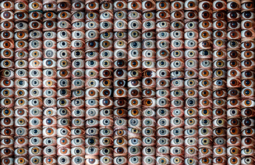
This gallery features images from the yearlong series of classes that I took in Ophthalmic Photography, as a part of the Biomedical Photographic Communications BS program at Rochester Institute of Technology. RIT is the only university in the USA to offer an Ophthalmic Photography program at an undergraduate level. This gallery also features images from my job as an Ophthalmic Photographer at the Virginia Eye Institute.
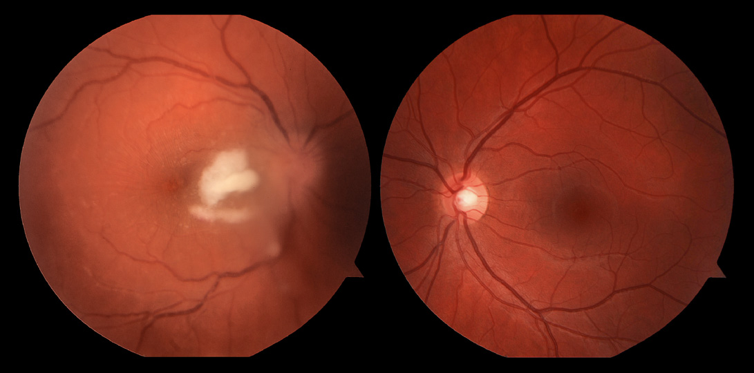
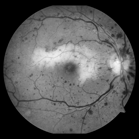
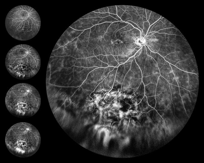
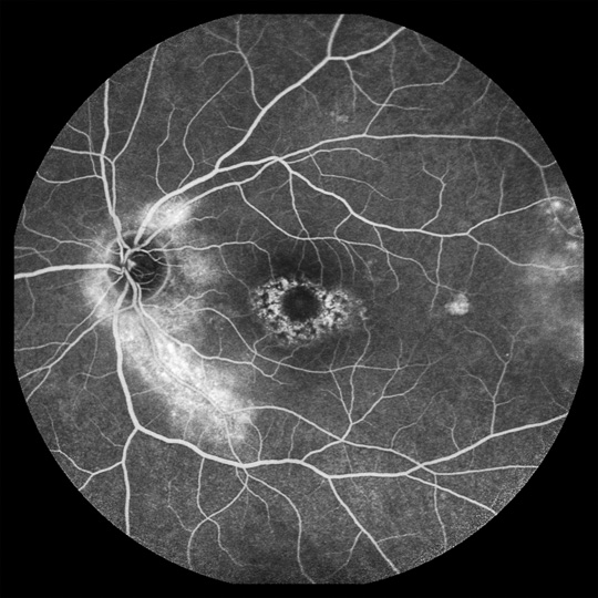
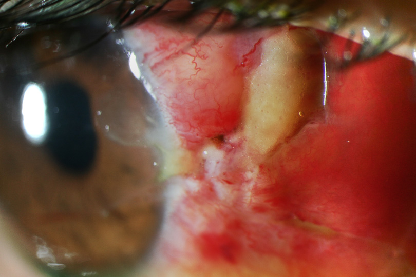
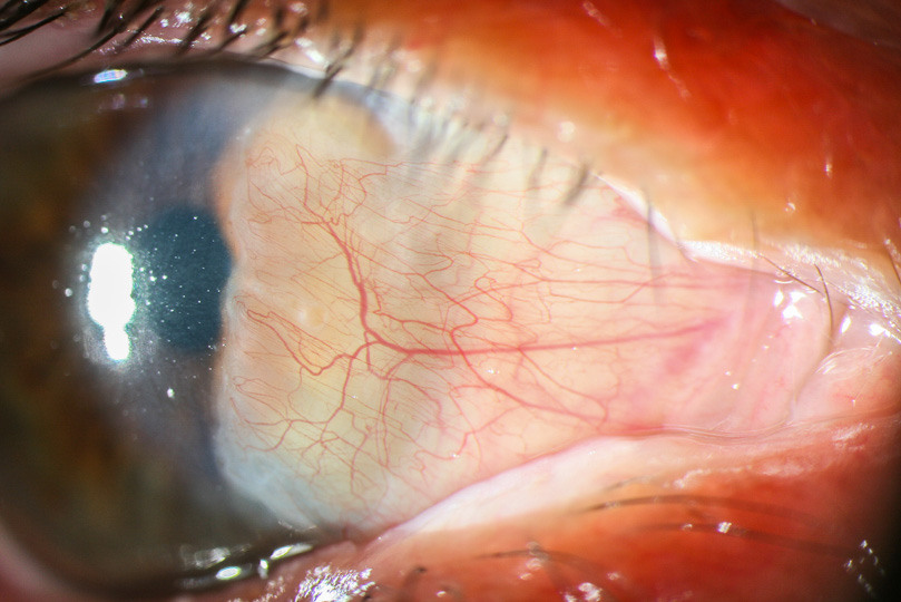
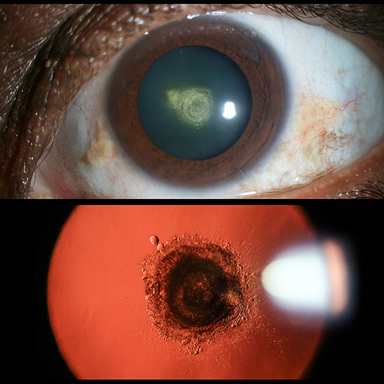
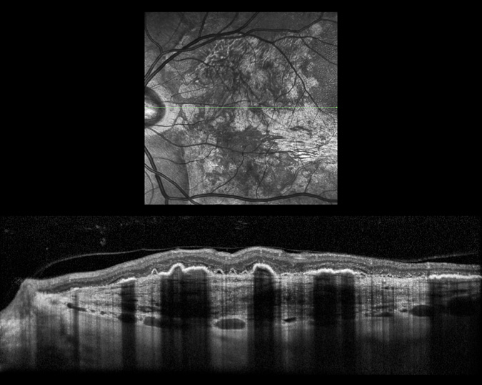
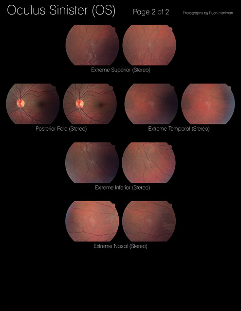
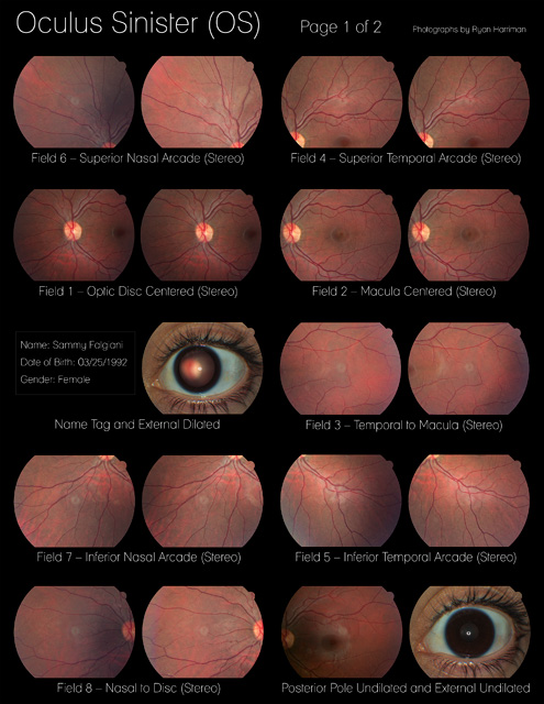
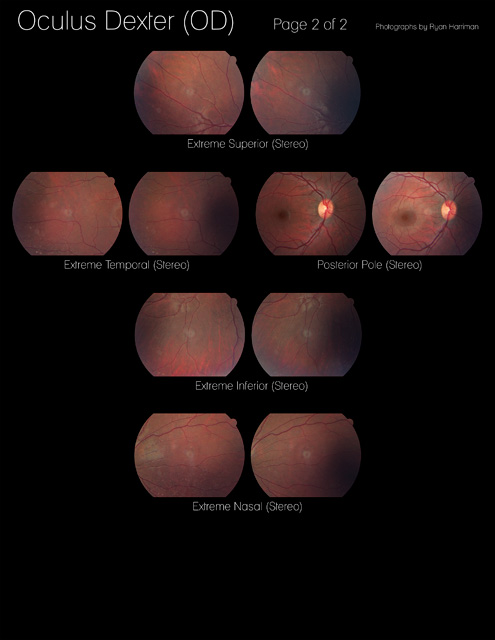
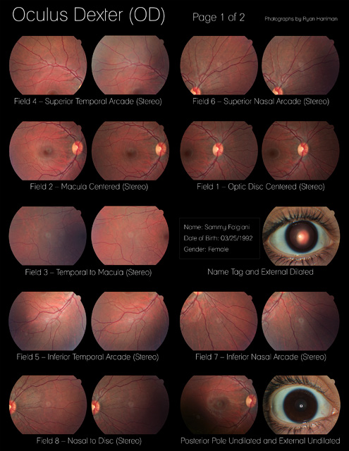
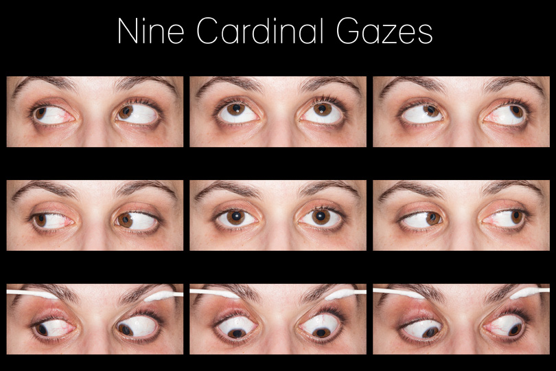
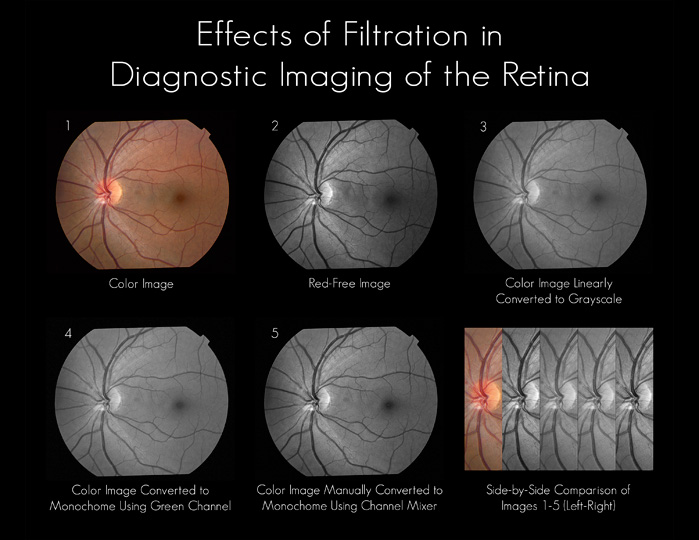
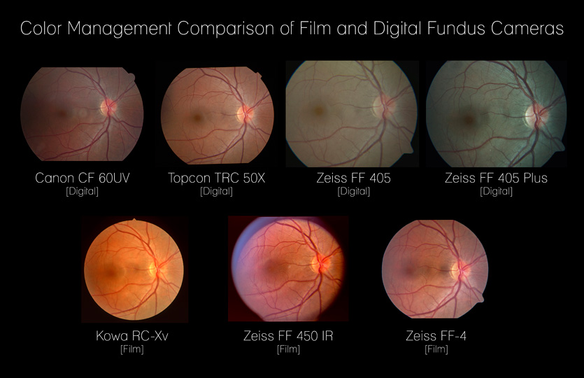
The Ophthalmic Photography classes taught the following skills:In-depth eye anatomy and physiologyIn-depth eye pathology (including diseases of the cornea, lens, vitreous, retina, choroid, optic nerve, etc.)Pharmacology (including classification, safety, and mechanism of action) of common ocular drugsExternal photography of the eyes with a DSLR (such as the “Nine Cardinal Gazes”)Fundus photography with digital and film fundus cameras, including Canon, Kowa, Topcon, and Zeiss modelsFluorescein Angiography (FA) and Indocyanine Green (ICG) imagingThe “Seven Standard Fields” and other variantsSpectral Domain Optical Coherence Tomography (SD-OCT) with the Heidelberg SpectralisAdvanced slit-lamp biomicroscopyTest protocol standards to participate in clinical studies
To view the "Image Flow" gallery, click here.